Back Of Neck Anatomy : Muscles Of Back Superficial Layers Superficial Muscles Posterior Neck And Back : The lumbar region of the spine, more commonly known as the lower back, is situated between the thoracic, or chest, region of the spine, and the sacrum.
bymamakingston•
0
Back Of Neck Anatomy : Muscles Of Back Superficial Layers Superficial Muscles Posterior Neck And Back : The lumbar region of the spine, more commonly known as the lower back, is situated between the thoracic, or chest, region of the spine, and the sacrum.. The hard white exterior covering of the tooth is the enamel. May 31, 2021 · the content of the neck is grouped into 4 neck spaces, called the compartments. Spinal anatomy is a remarkable combination of strong bones, flexible ligaments and tendons, large muscles and highly sensitive nerves. The back muscles stabilize and move the vertebral column, and are grouped according to the lengths and direction of the fascicles. They provide movements of the spine, stability to the trunk, as well as the coordination between the movements of the limbs and trunk.
The back muscles stabilize and move the vertebral column, and are grouped according to the lengths and direction of the fascicles. Still, many individuals pay far too little attention to them. Mar 16, 2020 · it runs from the neck to the upper back. Understanding the anatomy of your lower spine can help you communicate more effectively with the medical professionals who treat your lower back pain. The lumbar region of the spine, more commonly known as the lower back, is situated between the thoracic, or chest, region of the spine, and the sacrum.
Normal Anatomy Of The Deep Muscles Of The Back And Neck Medical Art Works from cdn.shopify.com As the tooth tapers below the gumline, the neck is formed. The hard white exterior covering of the tooth is the enamel. Jun 17, 2021 · back muscles. The inner portions of the tooth consist of the dentin, a bonelike tissue, and the pulp. Contains glands ( thyroid , parathyroid, and thymus ), the larynx , pharynx and trachea. The back muscles are divided into two large groups: The cervical spine supports the weight and movement of your head and protects the nerves exiting your brain. Neck, in land vertebrates, the portion of the body joining the head to the shoulders and chest.
The hard white exterior covering of the tooth is the enamel.
Understanding the anatomy of your lower spine can help you communicate more effectively with the medical professionals who treat your lower back pain. The muscles of the back are a group of strong, paired muscles that lie on the posterior aspect of the trunk. They provide movements of the spine, stability to the trunk, as well as the coordination between the movements of the limbs and trunk. Contains glands ( thyroid , parathyroid, and thymus ), the larynx , pharynx and trachea. The splenius muscles originate at the midline and run laterally and superiorly to their insertions. Apr 12, 2018 · the image below to shows all the major back muscles (as well as some neck muscles): Spinal anatomy is a remarkable combination of strong bones, flexible ligaments and tendons, large muscles and highly sensitive nerves. As the tooth tapers below the gumline, the neck is formed. From the sides and the back of the neck, the splenius capitis inserts onto the head region, and the splenius. The cervical spine protects the nerves connecting to the brain, allowing the head to move freely while supporting its weight. It is designed to be incredibly strong, protecting the highly sensitive nerve roots, yet highly flexible, providing for mobility on many different planes. The lumbar region of the spine, more commonly known as the lower back, is situated between the thoracic, or chest, region of the spine, and the sacrum. The inner portions of the tooth consist of the dentin, a bonelike tissue, and the pulp.
From the sides and the back of the neck, the splenius capitis inserts onto the head region, and the splenius. Some important structures contained in or passing through the neck include the seven cervical vertebrae and enclosed spinal cord, the jugular veins and carotid arteries, part of the esophagus, the larynx The back anatomy includes some of the most massive and functionally important muscles in the human body. Jun 17, 2021 · back muscles. It is designed to be incredibly strong, protecting the highly sensitive nerve roots, yet highly flexible, providing for mobility on many different planes.
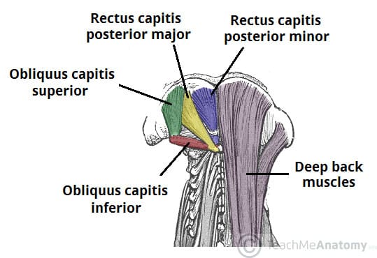
Muscles Of The Neck Teachmeanatomy from teachmeanatomy.info The rounded upper projections of the back teeth are cusps. The back muscles stabilize and move the vertebral column, and are grouped according to the lengths and direction of the fascicles. The cervical spine supports the weight and movement of your head and protects the nerves exiting your brain. Contains cervical vertebrae and postural muscles. Still, many individuals pay far too little attention to them. Understanding the anatomy of your lower spine can help you communicate more effectively with the medical professionals who treat your lower back pain. Mar 16, 2020 · it runs from the neck to the upper back. May 31, 2021 · the content of the neck is grouped into 4 neck spaces, called the compartments.
The cervical spine protects the nerves connecting to the brain, allowing the head to move freely while supporting its weight.
It consists of seven vertebrae. Jun 17, 2021 · back muscles. The muscles of the back are a group of strong, paired muscles that lie on the posterior aspect of the trunk. The rounded upper projections of the back teeth are cusps. The splenius muscles originate at the midline and run laterally and superiorly to their insertions. From the sides and the back of the neck, the splenius capitis inserts onto the head region, and the splenius. Mar 16, 2020 · it runs from the neck to the upper back. The back muscles are divided into two large groups: As the tooth tapers below the gumline, the neck is formed. Below the neck, holding the tooth into the bone, is the root of the tooth. Neck, in land vertebrates, the portion of the body joining the head to the shoulders and chest. The inner portions of the tooth consist of the dentin, a bonelike tissue, and the pulp. The hard white exterior covering of the tooth is the enamel.
The hard white exterior covering of the tooth is the enamel. Contains glands ( thyroid , parathyroid, and thymus ), the larynx , pharynx and trachea. The splenius muscles originate at the midline and run laterally and superiorly to their insertions. Some important structures contained in or passing through the neck include the seven cervical vertebrae and enclosed spinal cord, the jugular veins and carotid arteries, part of the esophagus, the larynx Still, many individuals pay far too little attention to them.
Upper Cervical Spine Disorders Anatomy Of The Head And Upper Neck from www.spineuniverse.com Apr 12, 2018 · the image below to shows all the major back muscles (as well as some neck muscles): The lumbar region of the spine, more commonly known as the lower back, is situated between the thoracic, or chest, region of the spine, and the sacrum. Spinal anatomy is a remarkable combination of strong bones, flexible ligaments and tendons, large muscles and highly sensitive nerves. It consists of seven vertebrae. The back muscles stabilize and move the vertebral column, and are grouped according to the lengths and direction of the fascicles. They provide movements of the spine, stability to the trunk, as well as the coordination between the movements of the limbs and trunk. Neck, in land vertebrates, the portion of the body joining the head to the shoulders and chest. As the tooth tapers below the gumline, the neck is formed.
Some important structures contained in or passing through the neck include the seven cervical vertebrae and enclosed spinal cord, the jugular veins and carotid arteries, part of the esophagus, the larynx
Still, many individuals pay far too little attention to them. Understanding the anatomy of your lower spine can help you communicate more effectively with the medical professionals who treat your lower back pain. The muscles of the back are a group of strong, paired muscles that lie on the posterior aspect of the trunk. The back muscles are divided into two large groups: It is designed to be incredibly strong, protecting the highly sensitive nerve roots, yet highly flexible, providing for mobility on many different planes. Jun 17, 2021 · back muscles. Contains cervical vertebrae and postural muscles. Spinal anatomy is a remarkable combination of strong bones, flexible ligaments and tendons, large muscles and highly sensitive nerves. The hard white exterior covering of the tooth is the enamel. As the tooth tapers below the gumline, the neck is formed. Mar 16, 2020 · it runs from the neck to the upper back. Apr 12, 2018 · the image below to shows all the major back muscles (as well as some neck muscles): The lumbar region of the spine, more commonly known as the lower back, is situated between the thoracic, or chest, region of the spine, and the sacrum.